Individual 3-D organ views and informationĭissection tools like pins, marker, scissors, scalpel, and forcepsĭetailed labels, information on frog classification, lifecycle, and organ functionsĪnatomical comparison of human and frog organs This is an excellent use of touch-driven educational activity that also happens to be odorless and environmentally sensitive. Frogs have been a long time model of understanding the function, position and relative size of organs, tissues, and systems. Once a student exposes the virtual organs, a simple click brings up a window that features a fully "spinnable" computer-generated image of the organ as well as an informative description.Įveryone is curious what the lies beyond the skin. Users can then follow the step-by-step instructions (with voice-over) that help them insert pins where needed and use a marker, scissors, scalpel and forceps to remove the frog's skin. Launch the app, and an upside-down frog appears on a blue field.

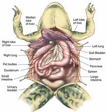
The large intestine which opens into a chamber called the cloaca.The frog dissection app should help win the day for those out to save the frogs. It is a fan like organ and lifting it up will reveal the small intestines. Mesentery - The mesentery holds together the small intestine. In fact, you might mistake them for intestines, and they sit on both the left and right side of the mesentery. Oviducts - The next noticeably large structure (in female frogs) is the oviducts. Locate the pancreas, which is a thin, flat, ribbon-like organ that lies between the stomach and the small intestine. Lift the small intestine to find the round, reddish spleen attached to the mesentery on the underside. Look for the duodenum, which is fairly straight, connecting to the rest of the intestine, which is coiled and connects to the mesentery. Gently pull out the stomach (but do not cut) to find:įind the small intestine which is connected to the stomach. To cut it out, you'll need to remove the liver first. Since frogs swallow their food whole, you can actually open the stomach to see what your frog ate. Stomach - Right underneath and curving below the liver is the stomach. The gall bladder which is a mall greenish-brown sac.In this case, the dye for the specimen makes two of the liver lobes dark blue. The frog liver has three lobes and sits to (your) right of the heart, almost on top of the stomach. Liver - One of the most noticeable structures in your open frog is the liver.You'll want to identify them first before cutting any of them out. There are several organs that sit 'on top' of other internal structures. If you don't get a dyed specimen, you will not see all the red and blue coloring. It should be noted here that these particular specimens are dyed to help students see the veins and blood vessels more clearly.

For comparison, the male frog will have no eggs, and you'll have a clear view of his abdominal cavity. In the picture above, the frog is a female, and you can see the eggs (the black, seed-like stuff). At this point, you should see some organs.
#Frog dissection diagram worksheet skin#
Remember to cut gently as you just want to peel the skin back and open the frog up. After you make the vertical incision, next you're going to want to peel the skin back on the ventral side of the frog by making two horizontal incisions - one up top by the frog's neck and one towards the bottom by its legs.

When you cut, you want to be careful to cut skin only so that you can peel away the layer gently. The first incision you make should be from the top of the frog's jaw all the way down to between his legs. Note you may have to break a bone to pin the frog at the hands so he's spread out.Ģ. Also, make sure insert your pins at an angle - putting them in straight up and down makes it easier for them to become dislodged while you're dissecting. It is easiest to pin the frog through the 'hand' and through the feet.


 0 kommentar(er)
